Medically reviewed by Benjamin M. Woodhead, DO
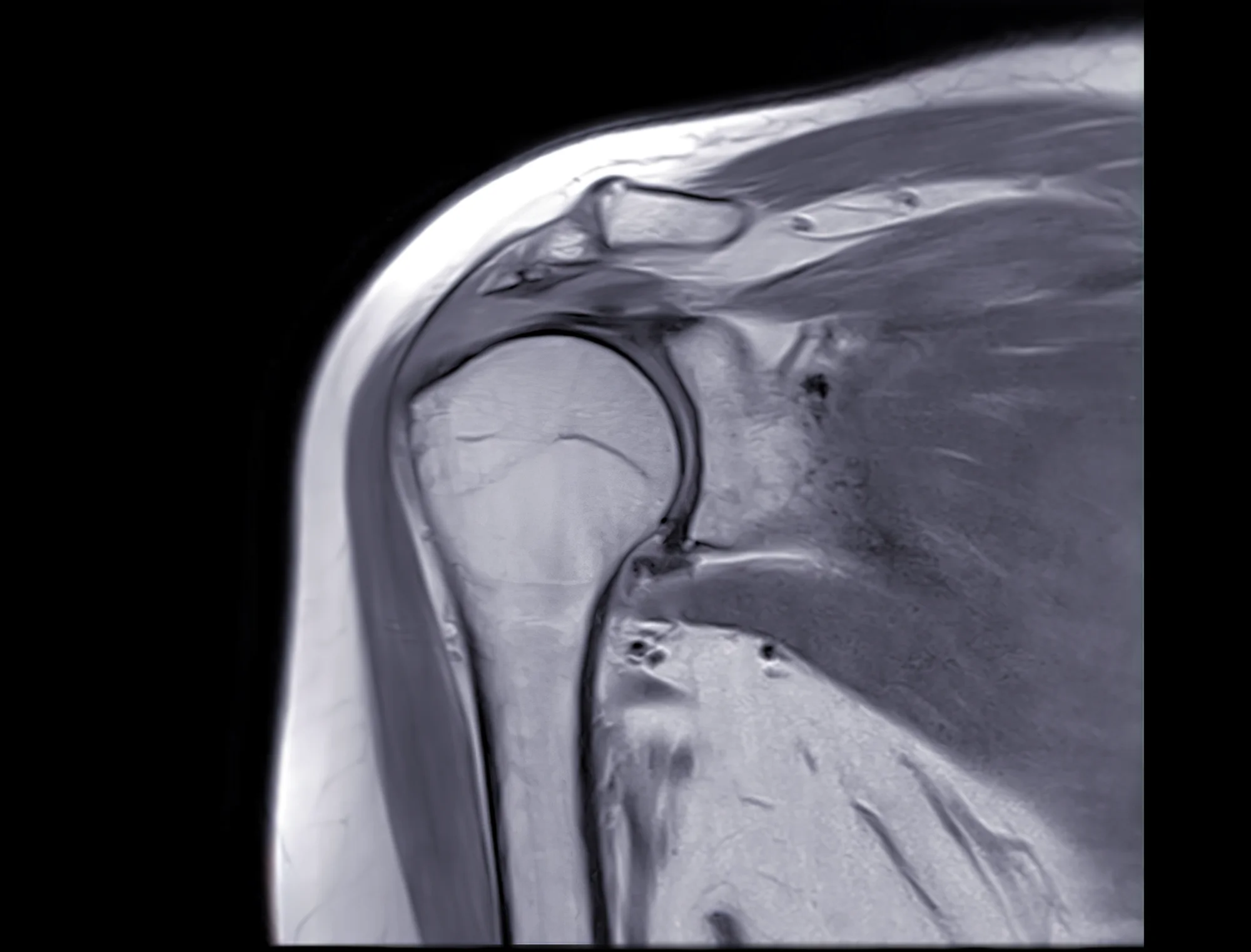
A shoulder MRI gives me a detailed look at structures I cannot fully evaluate on an X-ray or physical examination alone. I can see the rotator cuff, labrum, cartilage, tendons, and other soft tissues in much greater detail, which helps me understand what may be causing your symptoms. But an MRI is only one part of the evaluation. A scan can show a tear, degeneration, or other abnormality without telling me whether that finding is the source of your pain or whether it needs treatment. That is where your symptoms, physical examination, and the details of the MRI need to come together. In this post, I’ll explain what a shoulder MRI shows, what it cannot tell us by itself, and how I use those findings when evaluating patients in my Lincoln practice.
Key Takeaways
- An MRI maps tissue, not pain. Two people with nearly identical scans can feel completely different.
- It shows tendons, cartilage, labrum, muscle quality, and fluid very well. It shows bone detail less well than a CT scan.
- The scan is an adjunct to a physical exam, not a replacement for one. I read it alongside your history, never instead of it.
What a Shoulder MRI Is Actually Imaging
Magnetic resonance imaging (MRI) uses a strong magnetic field and radio waves to map water and fat inside tissue. No radiation is involved. Different tissues hold water differently, so tendon, muscle, cartilage, and fluid each return a different signal. The result is a stack of thin slices through the joint in several planes.
That water-mapping is the key to reading these scans sensibly. Damaged, inflamed, or degenerating tissue tends to hold more fluid, so it lights up. Bright signal means the tissue has changed, and nothing more specific than that. It does not tell anyone how much the change hurts.
What a Shoulder MRI Shows Well
Rotator cuff tendons are the most common reason I order one. MRI shows whether a tendon is torn, roughly how large the tear is, and how far the torn edge has retracted toward the shoulder blade. It is a strong test for ruling a full-thickness tear out.
It also shows muscle quality. When a cuff tendon has been torn for a long time, the muscle behind it thins and can fill in with fat. That finding influences whether a repair is likely to hold.
The labrum, the cartilage rim around the socket, images reasonably well. So do joint cartilage, fluid in the bursa, and swelling in the bone itself. A suspected labral tear is one of the situations where injecting contrast dye into the joint improves the read.
For bone shape and spurs, plain X-rays and CT still beat MRI.
What a Shoulder MRI Cannot Tell You
It cannot measure pain. This is the single most important limitation in my opinion. There is no sequence, no contrast agent, and no magnet strength that images suffering.
It cannot reliably date an injury. A tear from last month and a tear from four years ago can look strikingly similar. I can sometimes infer age from retraction and muscle changes, but that is an inference, not a measurement.
It cannot tell you whether you need surgery. That decision comes from your exam, your function, your goals, and your response to treatment so far. I have recommended repair for a modest-looking tear in a working farmer and watchful treatment for a larger one in someone comfortable with what their arm does.
Finally, it cannot separate a finding that matters from a finding that simply exists. Degenerative changes in the shoulder become steadily more common with age, and a meaningful share of people with no pain at all have imaging abnormalities in the rotator cuff. Your scan describes your shoulder. It does not diagnose your problem.
Words on Your Report That Sound Worse Than They Are
Tendinosis or tendinopathy means the tendon has degenerative change, not a tear. Signal change is the imaging equivalent of noticing that something looks different, with no claim about why. Fraying describes surface irregularity that is extremely common past middle age.
Bursitis means fluid in the bursa, which often accompanies impingement and usually responds to treatment. Mild glenohumeral or AC joint arthrosis describes wear that many adults have.
I would rather patients read these terms as descriptions of tissue than as verdicts on their future. That framing changes the whole conversation.
What I See in My Patients
I have patients that drive a long way to see me. They come to Lincoln from across southeast Nebraska, and when someone has spent ninety minutes in the truck, they usually want the scan done as soon as possible. I understand that, and while sometimes it is the right decision, often the better answer is an exam, an X-ray, and six weeks of conservative treatment first.
My fellowship in shoulder and elbow surgery was at the University of Washington, and that training along with my own experience left me with a firm bias toward the patient over the picture. In clinic, I see every week how loosely imaging findings line up with how people actually function. The gap is wider than most patients expect.
When I Order a Shoulder MRI, and When I Wait
I order one when the answer will change what we do. That usually means a suspected full-thickness cuff tear with weakness on exam, a shoulder that has not improved with a genuine course of therapy, a suspected labral or instability problem in a younger athlete, or planning before surgery.
I may wait when the exam and X-rays already tell the story, when the shoulder is early in a treatment plan that has not been given a fair trial, or when the likely findings would not change my recommendation. An early scan in that situation tends to produce anxiety rather than answers.
Sometimes I add contrast. A standard study images the shoulder as it sits, while an arthrogram involves injecting dye into the joint first to outline structures more sharply. I use it mainly for labral questions and for looking at a shoulder that has already had surgery. For most rotator cuff questions, a good standard shoulder MRI is enough.
Making the Scan Worth Having
A few practical things improve the result. Tell the imaging center about any metal in your body, including implants and older surgical hardware. Say something in advance if enclosed spaces bother you.
Hold still. Motion blurs the images more than people realize, and a repeated sequence adds time to a study you already want finished.
Bring the actual images to your appointment, not only the report. I want to look at the study myself rather than read someone else’s paragraph about it. If the scan lives on a disc or a patient portal, bring the disc or the login.
Summary
A shoulder MRI is excellent at showing tendon tears, muscle quality, labral injury, and fluid. It is poor at telling anyone how much a shoulder hurts, when an injury happened, or what should be done about it. Radiology terms like tendinosis, fraying, and mild arthrosis describe tissue rather than deliver a sentence. Findings grow more common with age, and plenty of them never cause a symptom.
If you already have a scan, do not spend another week interpreting it alone. Bring the images and the report to an appointment, and let us put them next to an exam and a conversation about what you actually need the arm to do. You can reach out to my office in Lincoln to get that started.
Frequently Asked Questions
Do I need an MRI for shoulder pain?
It depends, but usually not right away. Most shoulder pain gets evaluated well with a history, a physical exam, and X-rays, and many causes improve with treatment before imaging changes anything. I order a shoulder MRI when weakness, a suspected tear, or a failed course of therapy means the result would actually change the plan.
Does a shoulder MRI hurt?
The standard study is painless. You lie still while the machine works, and the main complaints are noise and the enclosed feeling. An arthrogram adds an injection of contrast into the joint beforehand, which is usually described as pressure rather than pain.
My report says I have a tear. Does that mean surgery?
No. A tear on imaging is one input among several. I weigh tear size, muscle quality, your strength on exam, how long symptoms have lasted, and what you need the shoulder to do. Many tears are managed well without an operation.
Is an ultrasound as good as an MRI?
For full-thickness rotator cuff tears in experienced hands, ultrasound can perform well and cost less. It is less reliable for partial tears and gives me less information for surgical planning, so MRI remains my usual choice when the findings will shape a decision.